


The life cycle and morphology of hantavirus represent a complex biological framework split between an asymptomatic persistent phase in rodent reservoirs and a destructive replication cycle in humans. Characterized by a pleomorphic, enveloped virion housing a trisegmented negative-sense RNA genome, understanding this structural design is crucial for combating severe spillover diseases like hantavirus pulmonary syndrome (HPS).
Life Cycle of Hantavirus
The hantavirus life cycle involves two distinct phases:
- Transmission /ecological cycle (in rodent reservoirs)
- Cellular replication cycle (within infected host cells, i.e., human).
Transmission & Ecological Cycle
Reservoir Hosts (Natural Life Cycle)
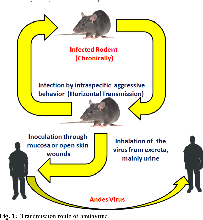
Hantaviruses are enzootic viruses that maintain persistent, lifelong infections in their rodent hosts.
Rodents (Primary Host)
deer mice, white-footed mice. rice rats, bank voles without causing apparent disease. Occurs primarily through aggressive behavior (biting), sexual contact, and maternal transmission (from mother to offspring)
Insectivores
shrews and moles (for some hantavirus species)
Viral Shedding
Infected rodents continuously shed virus in urine, droppings, and saliva.
Persistence
The virus persists in rodent populations through vertical transmission and horizontal transmission, ensuring the virus remains endemic.
What Is the Cellular Life Cycle of Hantavirus?
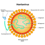
The virus is a tripartite, single-stranded RNA genome with three segments: L (large, polymerase), M (medium, glycoproteins), and S (small, nucleocapsid).
Attachment
Viral glycoproteins bind to host cell receptors, e.g., β₃ integrins.
Entry
Virus enters via endocytosis.
Uncoating
Viral envelope fuses with endosomal membrane in acidic endosomes/lysosomes, releasing nucleocapsid.
Transcription
Viral RNA-dependent RNA polymerase (RdRp) transcribes viral mRNA from genome RNA.
Translation
Host ribosomes translate viral proteins (nucleocapsid, glycoproteins, polymerase).
Replication
RdRp replicates the viral RNA genome in the cytoplasm.
Assembly
New virions assemble in the Golgi apparatus.
Egress
Mature virions exit via exocytosis through the cell membrane.
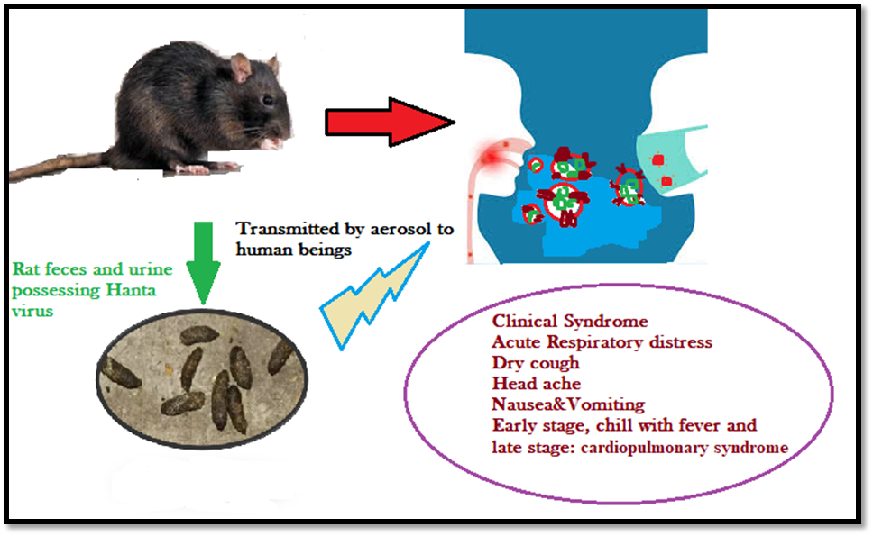
How Does Hantavirus Transmission and Human Infection Occur?
Primary Transmission Route
Inhalation of aerosolized virus from rodent urine, droppings, or saliva.
Less Common Routes
Direct contact with broken skin, mucous membranes, or rare rodent bites.
Human to Human
Only the Andes virus is known to spread person to person (close contact).
Incubation Period
1 to 2 weeks (up to 8 weeks in rare cases).
Disease Outcomes
Hantavirus Pulmonary Syndrome (HPS) in the Americas.
Hemorrhagic Fever with Renal Syndrome (HFRS) in Europe / Asia.
Key Distinction
The key distinction is that hantavirus causes asymptomatic persistent infection in rodents but severe, often fatal disease in humans upon spillover.
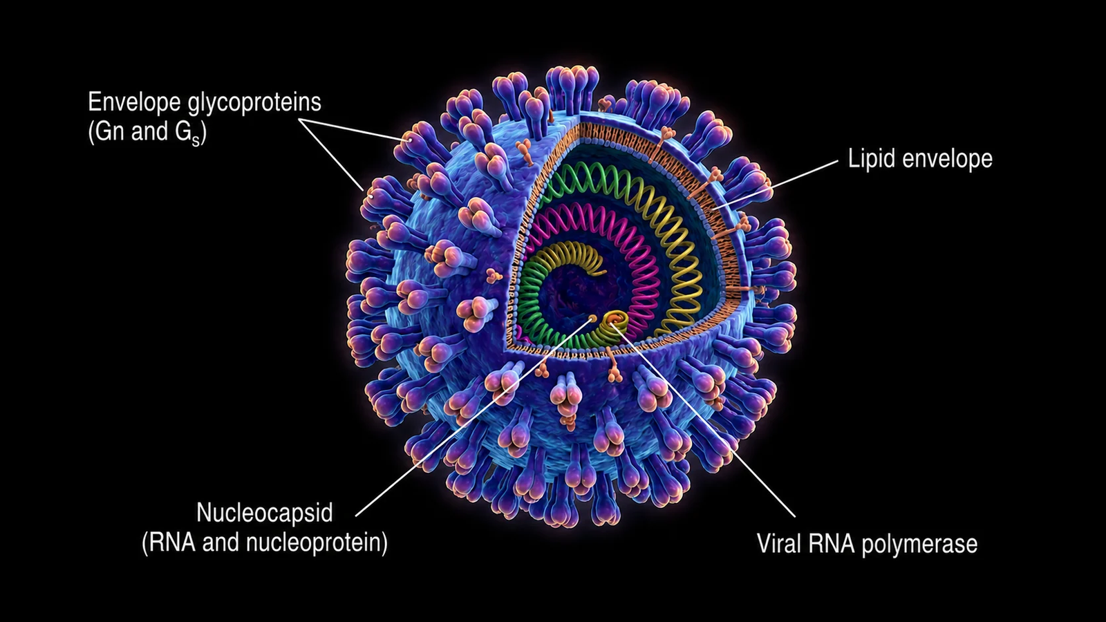
Morphological Study of Hantavirus
Hantavirus exhibits pleomorphic morphology with distinct structural features visible through electron microscopy.
Overall Virion Morphological Features
Shape
Spherical to pleomorphic, i.e., round, tubular, or irregular.
Diameter
88 to 160 nm (average ~120 nm to 135 nm).
Envelope
Single lipid bilayer, 5 nm thick.
Surface Projections
5 to 10 nm long spikes forming a square grid-like pattern.

Transmission Electron Micrograph of Sin Nombre Virus Particles Showing Spherical Orthohantavirus with Surface Spikes
Morphological Diversity (Cryo-EM Findings)
Recent cryo-electron microscopy revealed three distinct morphology classes that vary by virus strain.
Round
65% of old-world HTNV.
Tubular
30% of old-world HTNV.
Irregular
5% of old-world HTNV.
Black Creek Canal Virus (BCCV)
72% tubular with lengths up to 430 nm.
Sin Nombre Virus (MCV Strain)
53% irregular (highest among all strains).
What Are the Key Structural Components of a Hantavirus Virion?
Surface Structure
Glycoprotein Spikes
Tetramers of Gn/Gc glycoproteins extending ~10 nm from the lipid bilayer.
Square Grid-Like Pattern
Distinct from other Bunyavirales families.
Bare Patches
30 to 40 nm membrane areas devoid of spikes (more common in irregular particles).
Internal Structure
Ribonucleoproteins (RNPs)
Three rod-like structures (~10 nm thick) inside the virion.
Genome Segment Association
Each RNP contains one genome segment (S, M, or L) wrapped in nucleocapsid (N) protein.
RNP Configuration
RNPs appear as parallel straight rods or bent/curved projections toward the membrane.
Filamentous Ribonucleocapsid
200 to 300 nm long, 2 to 2.5 nm wide.
Genome Organization
| Segment | Size | Encodes Protein |
|---|---|---|
| L | 6.8 to 12 kb | RNA-dependent RNA polymerase (RdRp) |
| M | 3.2 to 4.9 kb | Glycoproteins Gn and Gc |
| S | 1 to 3 kb | N protein (nucleocapsid protein) |
Assembly and Building Sites
Primary Site
Endoplasmic reticulum-Golgi compartment.
Secondary Site
Host plasma membranes, especially for tubular particles.
Virion Release
Virions bud from Golgi and exit via exocytosis.
The morphological diversity (round, tubular, irregular) suggests pleiotropic assembly without a matrix protein, and the functional significance of different morphologies for viral entry remains under investigation.
Difference Between Key Structures of Hantavirus and Andes Hantavirus
Hantavirus and Andes hantavirus have the same basic virion architecture: they are enveloped, roughly spherical viruses with a segmented negative-sense RNA genome made of L, M, and S segments. The main structural difference is that Andes virus has more recently characterized, high-resolution glycoprotein and nucleoprotein structures that show detailed features of its surface spikes and RNA-binding protein organization.
Shared Core Structure
Both belong to the hantavirus family and share the same overall plan:
- A lipid envelope.
- Surface glycoproteins Gn and Gc.
- A tripartite RNA genome: L, M, and S segments.
- A nucleoprotein that packages the RNA genome.
Andes-Specific Structural Features
Andes virus is a specific hantavirus, so it does not have a completely different virus body plan. What stands out is the detailed structure of its proteins:
- Its nucleoprotein core has an N lobe and C lobe that form an RNA-binding crevice.
- Its glycoproteins form a membrane-embedded tetramer, with newer structural studies featured in the National Institutes of Health (NIH) database showing pH-sensing and organization details important for cell entry.
Difference Between Them
So the difference is not “hantavirus vs. Andes hantavirus” at the level of broad virus shape; Andes virus is one member of the hantavirus group. The real difference is in species-specific protein structure, especially the Andes virus glycoprotein tetramer and nucleoprotein architecture, which have been solved in higher detail and are linked to its unique biology and person-to-person spread.
Simple Way to Think About It
Think of hantavirus as the family blueprint and Andes virus as one model built from that blueprint. The frame is similar, but the exact arrangement of the surface proteins and internal RNA-binding proteins differs enough to affect infectivity, immune recognition, and transmission.
FAQ
Q: What are the three genome segments of Hantavirus?
The hantavirus genome consists of three negative-sense single-stranded RNA segments: the Large (L) segment encoding the RNA-dependent RNA polymerase, the Medium (M) segment encoding the surface glycoproteins (Gn and Gc), and the Small (S) segment encoding the nucleocapsid protein.
Q: How does the morphology of Andes hantavirus differ from other strains?
While sharing the same basic enveloped, trisegmented structure, Andes virus exhibits unique species-specific protein architecture in its nucleoprotein lobes and surface glycoprotein tetramers, which are directly associated with its ability to spread via person-to-person transmission.
1 Comment
Thottpalayam virus (TMPV): 7 Vital Facts on This Unique Pathogen
June 11, 2026[…] virus is a unique, shrew-borne hantavirus discovered in India in 1964. While human infection is confirmed serologically, clinical disease […]