

When examining the Key Structure of Hantavirus, these pathogens are identified as enveloped, negative-sense RNA viruses characterized by a unique trisegmented genome and a distinct surface glycoprotein lattice. Understanding the molecular architecture of its L, M, and S segments, alongside the fusion mechanisms of its Gn and Gc proteins, is critical for understanding viral entry and developing targeted vaccine designs.

What is the genomic structure of hantavirus?
Trisegmented, single-stranded, negative-sense RNA divided into three segments:
- S segment (1 to 3 kb): encodes the nucleocapsid (N) protein
- M segment (3.2 to 4.9 kb): encodes two glycoproteins (Gn and Gc) as a polyprotein precursor
- L segment (6.8 to 12 kb): encodes the RNA-dependent RNA polymerase (L protein)
What Are the Key Structural Proteins of Hantavirus?
| Protein | Function |
| N (nucleocapsid) protein | Encapsidates viral RNA to form ribonucleoproteins (RNPs); protects genome |
| Gn&Gc glycoproteins | Form heterodimers that assemble into tetrameric spikes on the surface; mediate virus attachment and entry via receptor-mediated endocytosis and membrane fusion |
| L protein | RNA-dependent RNA polymerase (RdRp), responsible for transcription and replication, performs cap-snatching to prime mRNA synthesis. |
What Does the Hantavirus Virion Architecture Look Like?
- Enveloped, spherical to pleomorphic particles
- Diameter: 80 to 160 nm, typically 120 to 160 nm
- Lipid envelope: derived from host Golgi apparatus
- The surface displays a characteristic square lattice formed by Gn-Gc spike tetramers.
Internal ribonucleoprotein complexes (RNA + N protein) enclosed within the envelope
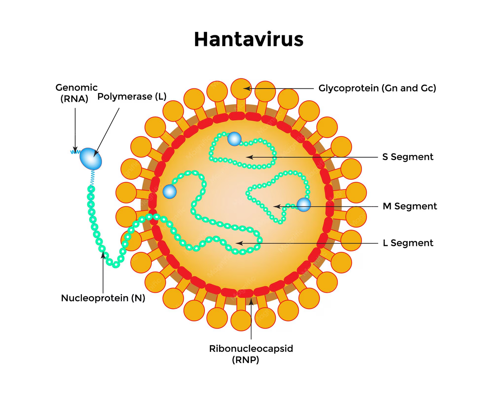
Schematic of Hantavirus structure showing L/M/S RNA segments, Gn/Gc glycoprotein spikes ,lipid envelope and ribonucleocapsid
Transmission electron micrograph of Sin Nombre virus particles.
Transmission electron micrograph of Sin Nombre virus particles showing roughly spherical virions with a textured outer layer. The Gn and Gc glycoproteins are the sole targets of neutralizing antibodies, making them critical for vaccine design.
How do Gn and Gc protein interactions facilitate host cell entry?
According to established structural biology data maintained by organizations like the National Institutes of Health (NIH), the Gn-Gc protein interaction facilitates host cell entry through a coordinated multi-step process:
Receptor Binding (Primarily Gn)
Gn (glycoprotein N-terminal ) mediates attachment to host cell receptors.
- Primary receptors: β₃ integrins (e.g., αvβ₃, α5β1)
- Alternative receptors: protocadherin-1 (PCDH1) and DC-SIGN
- Gn exposes receptor-binding domains that recognize and bind these cell surface proteins, anchoring the virion to the host cell.
Internalization via Endocytosis
- After receptor binding, the virus is internalized via receptor-mediated endocytosis into endosomal compartments.
- The exact mechanism of endocytosis varies by hantavirus species but typically involves clathrin-mediated pathways.
Low pH-Triggered Conformational Change (gc-driven)
- As the endosome matures, pH drops to ~5.0 to 6.0
- This acidic environment triggers Gc (glycoprotein C) to undergo a dramatic conformational rearrangement.
- Gc adopts a class II fusion protein fold.
Membrane Fusion (GC-mediated)
- Gc is the fusogen: it drives the fusion of viral and endosomal membranes.
- The conformational change exposes a hydrophobic fusion peptide in Gc that inserts into the endosomal membrane.
- Gc then refolds into a post-fusion hairpin structure, pulling the viral and host membranes together
- This fusion creates a pore through which the viral ribonulceocapsid (RNP) is released into the cytoplasm.
Gn-Gc Heterodimer Stability
- This heterodimeric arrangement is essential for proper folding, transport to the Golgi, and maintaining the fusion-competent state of Gc until low pH is encountered.
- Gn and Gc form stable heterodimers that assemble into tetrameric spikes on the virion surface.
| Step | Primary Protein | Function |
| Attachment | Gn | Binds β₃ integrins / PCDH1 receptors on host cell |
| Endocytosis | Both | Virus internalized into endosome |
| pH sensing | Gc | Acidic pH triggers conformational change. |
| Membrane fusion | Gc | Fusion peptide inserts: hairpin formation drives fusion |
| RNPs release | Both | Viral genome released into cytoplasm |
The Gn-Gc interaction is therefore essential: Gn positions the virus at the cell surface, while Gc executes the energetically demanding membrane fusion that delivers the viral genome into the host cell
FAQ – Key Structure of Hantavirus
Q: What are the three RNA segments of the Hantavirus genome?
he hantavirus genome is trisegmented into the Large (L) segment, which encodes viral polymerase; the Medium (M) segment, which encodes surface glycoproteins; and the Small (S) segment, which encodes the nucleocapsid protein.
Q: How does Hantavirus enter host cells?
Hantavirus enters cells through a multi-step process where the Gn protein binds to host surface receptors (like β₃ integrins) and the Gc protein mediates low-pH-triggered membrane fusion inside the endosome.