



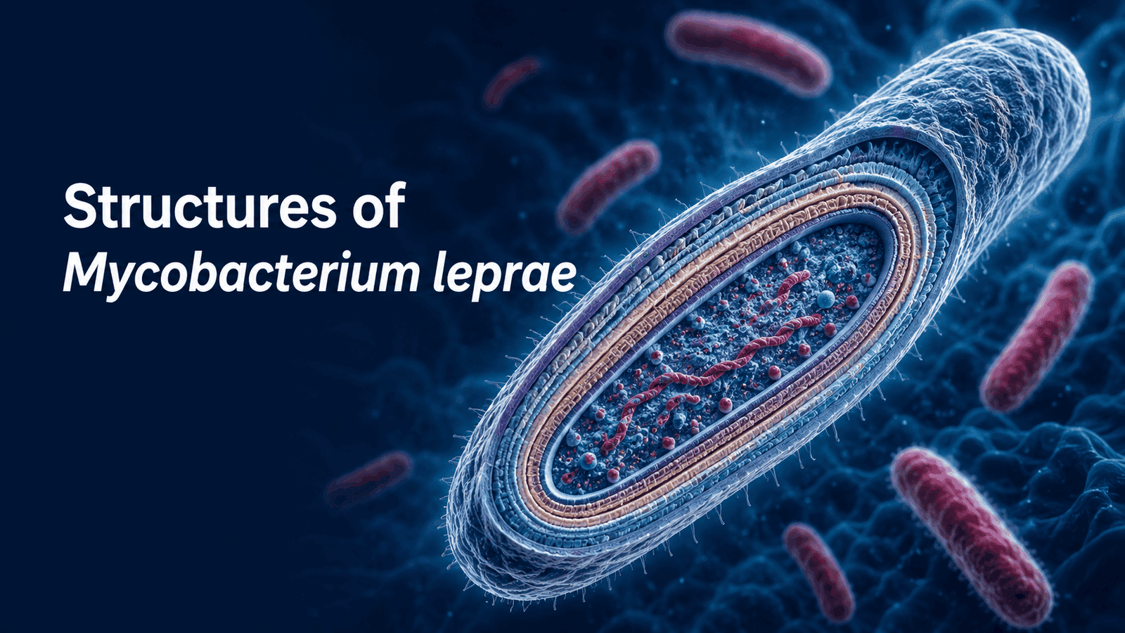
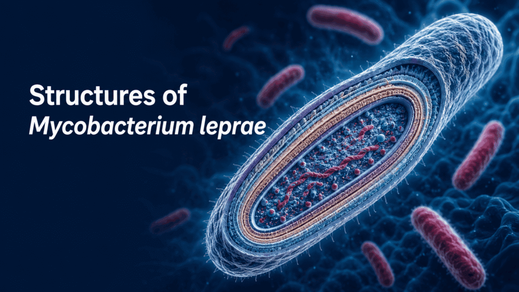
Mycobacterium leprae (the causative agent of leprosy/Hansen’s disease) is a straight or slightly curved, slender, acid-fast bacillus with a distinctive multilayered cell envelope and several structural adaptations for intracellular survival.
Gross morphology and basic features
- Shape and size: straight or slightly curved rods, about 1 to 8 micrometers in length and 0.2 to 0.5 micrometers in width.
- Arrangement: often appear fragmented or beaded; in tissues they may lie in parallel rows, giving a “cigar-bundle” appearance due to being bound by lipid-like substances (glia).
- Staining: acid-fast (retain carbolfuchsin despite acid-alcohol decolorization), but less resistant to strong acid than M. tuberculosis, typically described as resistant to about 5% sulphuric acid.
- Motility and spores: non-motile, non-sporing, and capsulated.
- Division: by binary fission; live bacilli stain brightly and uniformly (solid). While altered/dead bacilli stain weakly and granularly on Ziehl-Neelsen-type stains.
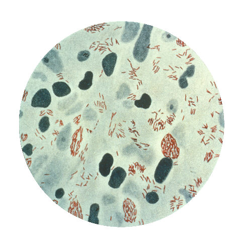
Mycobacterium leprae in lepromatous leprosy, seen by modified AFB Wade Fite staining at 100X magnification.
Microscopic view of numerous red, rod-shaped acid-fast bacilli of M. leprae in tissue (Wade-Fite stain).
Ultrastructure: major components
Ultrastructurally, M. leprae has four principal components: capsule, cell wall, cell membrane, and cytoplasm.
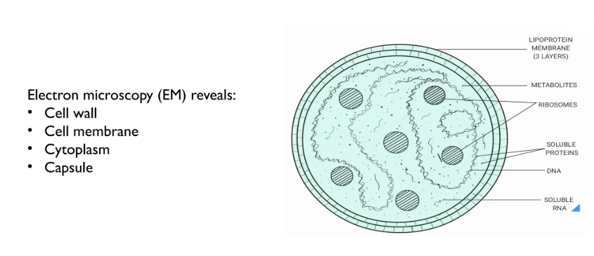
1. Capsule:
- Described as an electron-transparent zone of foamy or vesicular material surrounding the cell.
- Contains characteristic capsular lipids, notably:
- Phthiocerol dimycocerosates (PDIM).
- Phenolic glycolipid-1 (PGL-1)
- Function: Protects the bacillus from lysosomal enzymes and host metabolites.
- PGL-1 and related lipids are released into host fluids and are used in serological diagnosis (e.g., anti-PGL-1 assays).
2. Cell Wall:
- The cell wall of M. leprae is complex and critical for its acid-fastness and immune interactions. Sources describe it in slightly different but compatible ways:
- Thickness: about 20 nm.
- General architecture: An inner layer rich in peptidoglycan (electron-dense).
- An outer layer containing lipids and lipid-protein complexes, including lipopolysaccharide-like structures (electron-lucent).
- Biochemical model (often used for mycobacteria and specifically cited for M. leprae).
- Innermost: peptidoglycan–provides shape and rigidity.
- Attached to it: lipoarabinomannan-B (LAM-B)—highly immunogenetic; used in serodiagnosis.
- Then: a mycolic acid layer—long-chain fatty acids that confer acid fastness and impermeability.
- Outermost: phenolic glycolipid-1 (PGL-1)—contributes to virulence and immune modulation; also used in serology.
- Functional roles:
- Mediates nutrient uptake.
- Cell-wall proteins are major targets of T-cell immunity.
- It is the last structure to disappear during effective chemotherapy, which is why bacilli may remain visible for some time after treatment initiation.
3. Cell membrane:
- Lies beneath the cell wall and is composed of lipids (phospholipids) and proteins.
- Key proteins include MMP-I and MMP-II (mycobacterial membrane proteins)
1. Functions:
- Controls transport of molecules into and out of the organism.
- Houses enzymes and transport systems essential for metabolism and survival within macrophages and Schwann cells.
4. Cytoplasm:
1) Contains:
- DNA and RNA (genetic material and transcription/translation machinery).
- Storage granules (e.g., for lipids and other metabolites).
2) Functional significance:
- Site of protein synthesis, replication, and metabolic activity.
- Gel electrophoresis of cytoplasmic proteins shows several major proteins, reflecting its streamlined but specialized metabolism as an obligate intracellular pathogen.
Structural adaptations linked to pathogenicity:
- Obligate intracellular lifestyle: M. leprae parasitizes macrophages (histocytes) and Schwann cells in peripheral nerves; its structure supports survival in these niches.
- Reduced genome and metabolic capacity: many genes required for independent growth are lost, which is one reason it cannot be cultured in artificial media and must be grown in armadillo footpads or certain animal models.
- Lipid-rich envelope (mycolic acids, PDIM, PGL-1, LAM):
- Confers acid-fastness
- Provides resistance to host-killing mechanisms.
- Modulates host immunity (e.g., PGL-1 can suppress cellular immune responses).