



1. Life Cycle Of Ascaris lumbricoides
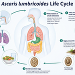
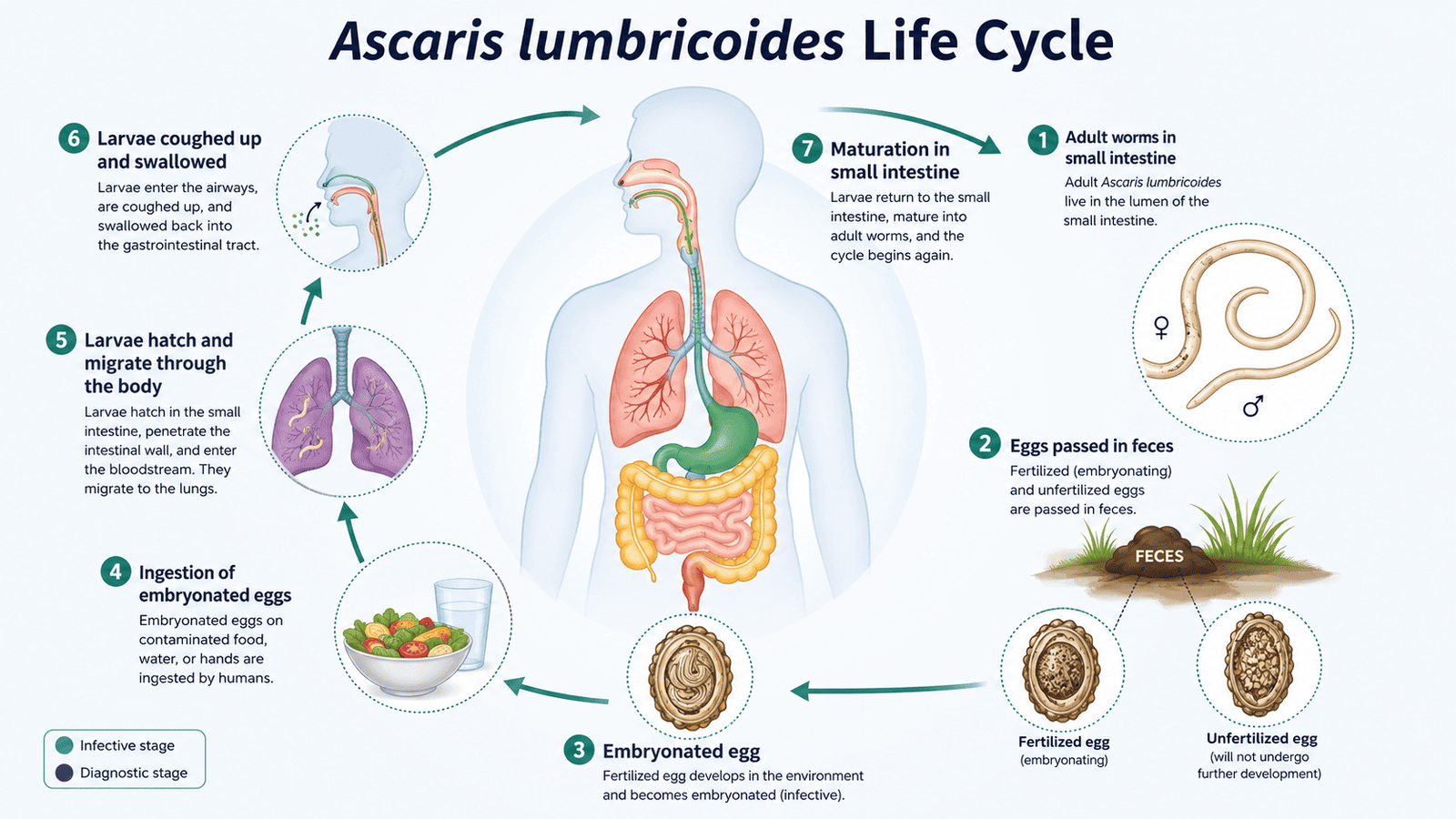
The life cycle of Ascaris lumbricoides is completed in a single human host, with eggs developing in the external environment, i.e., in soil.
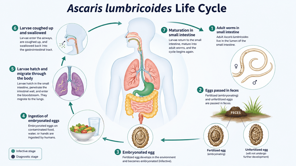
Ascariasis Life Cycle
Stages of the Life Cycle:
1. Eggs in faces (non-infective stage):
- Adult female worms in the small intestine produce up to 2 lakh to 2.5 lakh eggs per day.
- Eggs are passed in feces in an unembryonated (non-infective) form.
2. Development in soil (embryonation):
- Under favorable conditions (worm temperature is ~20 to ~25°C, moisture, and oxygen), eggs embryonate in soil over 3 to 6 weeks (commonly ~4 weeks).
- A second-stage (rhabditiform) larva develops inside the egg; this is the infective stage.
3. Ingestion of infective eggs (human infection):
- Humans become infected by ingesting embryonated eggs via contaminated food, water, soil, or unwashed hands.
- In the small intestine, the alkaline pH and bile stimulate the egg to hatch, releasing larvae.
4. Larval migration through tissues:
- Larvae penetrate the intestinal wall and enter the portal circulation, reaching the liver, then the heart, and then the lungs (within about 4 to 7 days).
- In the lungs, larvae:
→ enter alveoli,
→ Molt twice and grow,
→ Remain for about 1 to 2 weeks.
5. Return to the gastrointestinal tract:
- Larvae ascend the bronchi→ trachea→pharynx, are coughed up, and then swallowed.
- They pass through the esophagus and stomach back to the small intestine.
6. Maturation to adult worms:
- In the small intestine (mainly jejunum), larvae undergo two more molts and mature into adult worms.
- Sexual maturation occurs within 6 to 10 weeks after infection
- Adult males and females mate; females begin laying eggs, which are excreted in feces, continuing the cycle.
- Total time from ingestion of eggs to stool appearance of new eggs in stool is about 9 to 11 weeks.
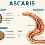
2. Morphology Of Ascaris lumbricoides
Adult worm:
Ascaris lumbricoides is the largest intestinal nematode infecting humans. resembling an earthworm in appearance. The sexes are separated and show marked sexual dimorphism, with the female generally larger than the male.
1. General features:
- Shape: Elongated, cylindrical, smooth, and tapering at both ends; the anterior end is thinner than the posterior.
- Size: Females are usually longer and thicker than males.
- Surface: finely striated, thick cuticle
- Colour: creamy white to pinkish when worms are fresh; may turn whitish after excretion.
- Mouth: Located at the anterior end and surrounded by three lips—one dorsal and two ventrolateral
- Digestive tract: Complete, consisting of mouth, pharynx, intestine, and anus.
- Esophagus: club-shaped, muscular.
- Habitat: Adult worms live mainly in the lumen of the human small intestine.
- Sexes: Male and female worms are separate individuals.
- Body covering: A tough, noncellular cuticle with fine transverse striations.
2. Sexual dimorphism:
Male: body smaller
→ Length: about 15 to 30 cm (commonly ~ 25cm)
→ Diameter: ~ 3 to 4 mm.
→ Posterior end: curved ventrally (hook-like).
→ Reproductive system: single set of genitalia with two small, equal spicules used in copulation.
→ A pair of needle-like penial setae in cloaca
→ prenatal and postnatal papillae present to help in copulation.
→ Function: Sperm production and transfer
Female: body larger
→ length: about 20 to 40 cm (commonly ~ 30 to 35 cm).
→Diameter: ~ 6 to 8 mm.
→ Posterior end: straight
—>Reproductive system: Anus and genital pore are separate. Genital pore midventral at about ⅓ body length behind anterior end.
—> No penial setae.
→ prenatal and postnatal papillae absent
→ Function: Egg production.
image
Thus, the most useful external distinctions are the smaller size and curved tail of the male versus the larger size, straight tail, and ventral vulva of the female.