



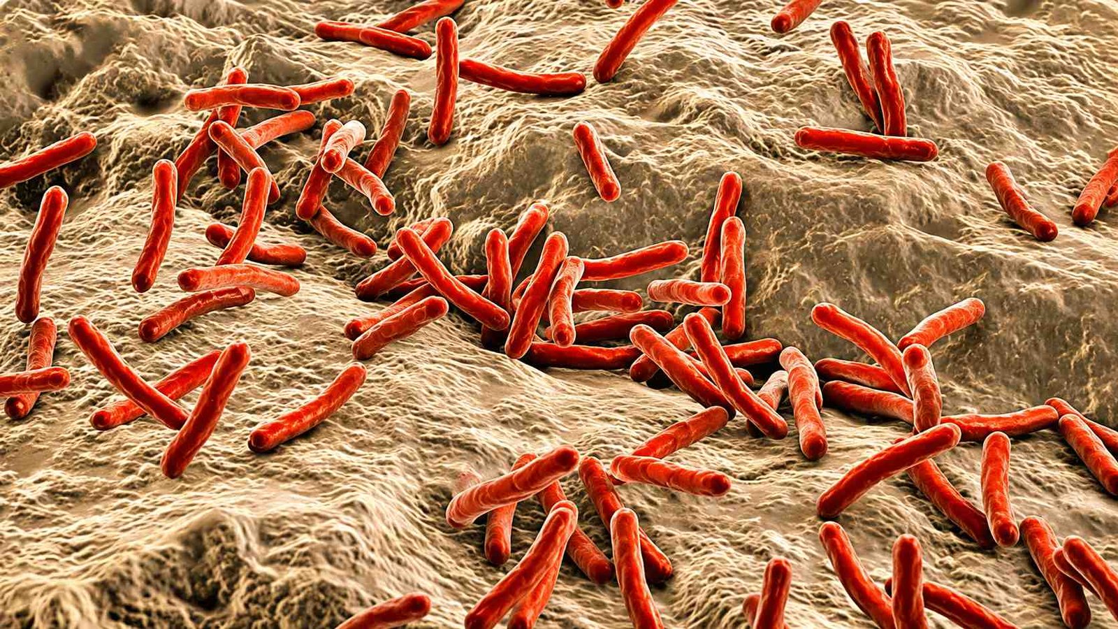
Mycobacterium leprae is a slender, rod-shaped bacterium. acid-fast bacillus with distinctive morphological features that aid its identification in clinical specimens and differentiate it from other mycobacteria.
📌 Key Takeaways: Morphology & Tropism of Mycobacterium leprae
- Morphological Characteristics: Slender, acid-fast rod (1–8 µm × 0.2–0.5 µm); non-motile, non-sporing; often arranged in parallel “cigar-packet” bundles or clumps (globi).
- Cell Wall Structure: Lipid-rich outer layer containing mycolic acids, conferring acid-fastness (stains bright red via Ziehl-Neelsen/Fite-Faraco stains).
- Thermotropism (Cooler Preference): Prefers lower temperatures (27°C–30°C), selectively colonizing peripheral skin, cool nerve endings, nasal mucosa, and testes.
- Schwann Cell Tropism: Uniquely binds and invades Schwann cells in peripheral nerves, causing demyelination, sensory loss, and secondary deformities.
General Morphological Features:
- Shape: Slender, straight, or slightly curved rods with parallel sides and rounded ends.
- Size: 1 to 8 micrometers in length and 0.2 to 0.5 micrometers in width (diameter ~ 0.3 micrometers).
- Arrangement: Singly, in parallel bundles (“cigar packet” or “cigar bundle” appearance), or in clumps called globi inside macrophages (lepra cells). In tissue smears, bacilli may appear as single rods, within phagocytes, or in spheroidal clumps.
- Motility and Spores: Non-motile and non-sporing.
- Capsule: Classically described as non-capsulated, though some sources note a slimy surface layer (G-substance) aiding adhesion.
- Gram reaction: Gram-positive cell wall (peptidoglycan present) but stains poorly with ordinary Gram stains; best visualized with acid-fast stains.
Acid-Fast Nature and Staining Characteristics:
M. leprae is acid-fast due to a cell wall rich in mycolic acids and other complex lipids, which resist decolorization by acid-alcohol after staining with carbol fuchsin.
Ziehl-Neelsen (ZN) / Fite-Faraco stain:
Bacilli stain bright red against a blue/green background.
Viability assessment in smears:
- Viable bacilli: Uniformly and solidly stained, bright red rods.
- Non-viable/degenerate bacilli: fragmented, granular, irregularly stained.
Metachromatic granules:
Bead-like thickening may be seen along the bacillus, giving a “beaded” appearance in some preparations.
Cell-wall and structural details:
Ultrastructurally. M. leprae has a typical mycobacterial cell envelope:
Cell wall:
~ 15 to 20 nm thick, composed of an inner peptidoglycan layer and an outer lipid-rich layer containing mycolic acids, arabinogalactan, and other glycolipids.
Waxy coating:
Thick, lipid-rich outer layer responsible for
- Acid-fastness
- Resistance to many disinfectants and host defenses.
- Poor uptake of routine stains.
Cell membrane:
Underlines the cell wall; cytoplasm contains ribosomes and nucleoid but no well-defined organelles (typical of prokaryotes).
Morphological Variability and Pleomorphism:
M. leprae is pleomorphic, showing variation in shape and arrangement depending on the following:
- Host immune status, e.g., numerous bacilli in globes in lepromatous leprosy vs. few in tuberculoid forms.
- Stages of infections and tissue type (skin, nerve, macrophages).
- Degenerative changes: cubical, lateral, or branching forms may be observed, especially in older or dying bacilli.
Key Diagnostic Morphological Points:
- Slender, straight/slightly curved rods, 1-8 * 0.2-0.5 μm.
- Parallel sides, rounded ends, “cigar packet” bundles, and globi in macrophages.
- Acid-fast (ZN/Fite stain): bright red rods, viable=solid & uniform; dead=fragmented and granular.
- Non-motile, non-sporing, Gram-positive cell wall visualized mainly by acid-fast stains.
- Pleomorphic; beaded forms and metachromatic granules may be seen.
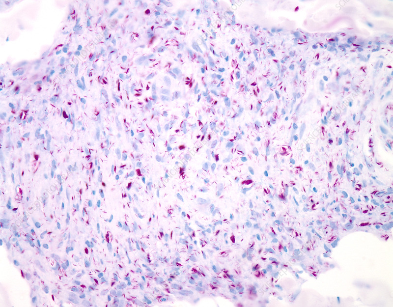
Mycobacterium leprae in lepromatous leprosy, seen by modified AFB Wade Fite staining at 100X magnification.
These morphological features, together with clinical findings and epidemiology, support the diagnosis of leprosy and help distinguish M. leprae from other acid-fast bacilli such as M. tuberculosis.
Sites of Mycobacterium leprae Infection and the Reasons:
Leprosy bacteria (Mycobacterium leprae) are mostly present in the skin and peripheral nerves, with additional involvement of the mucosa of the upper respiratory tract, eyes, and testes.
Primary sites of infection:
The disease predominantly affects:
- Skin: causing characteristic lesions, patches, nodules, and thickening.
- Peripheral nerves: leading to loss of sensation, muscle weakness, and deformities.
- Upper respiratory mucosa (nose and mouth): the main route of bacterial entry and exit.
- Eyes and testes: especially in cooler body regions.
Temperature preference (thermotropism):
M. leprae is an obligate intracellular bacillus that grows best at cooler temperatures (approximately 27 to 30°C), which are found in superficial tissues rather than at the core body temperature (37°C). This explains why it colonizes:
- Skin (especially extremities like ears, nose, fingers, and toes)
- Peripheral nerves in limbs
- Anterior chambers of the eye
- Testes (which are outside the main body cavity and cooler)
This is also why armadillos, which have lower body temperatures than most mammals, can serve as natural reservoirs.
Specific cellular binding:
M. laprae has a unique ability to bind to Schwann cells (the supporting cells of peripheral nerves). The bacterium expresses a trisaccharide on its surface that binds specifically to the basal lamina of Schwann cells, making it the only known bacterium that actively invades peripheral nerves. This nerve invasion leads to the following:
- Demyelination and nerve damage
- Loss of sensation (anesthesia)
- Secondary injuries and deformities due to lack of protective sensation
Clinical significance:
Because the bacteria concentrate in skin and nerves, leprosy manifests primarily as
- Hypopigmented or reddish skin patches with reduced sensation.
- Thickened peripheral nerves (e.g., ulnar, median, posterior tibial)
- Nerve function impairment (loss of pain, temperature, and touch sensation)
The coolest place in the human body:
The coolest parts of the human body under normal conditions are the extremities and appendages, particularly the following:
- Fingers and toes, especially the tips
- Ears including the earlobes
- Tip of the nose
These areas can have skin temperatures as low as 24°C to 30°C (75 to 86°F), compared to the core body temperature of about 37°C (98.6°F).
Why are these areas the coolest?
- Distance from the heart: The extremities are farthest from the heart, so blood reaching them has already passed through extensive vascular networks, losing heat along the way.
- Reduced blood flow in cold conditions: The body prioritizes maintaining core temperature (vital organs like brain, heart, and lungs) over peripheral temperature. When exposed to cold, blood vessels in the skin and extremities constrict (vasoconstriction), reducing blood flow and heat loss from these areas.
- High surface-area-to-volume ratio: Fingers, toes, ears, and the nose have relatively large surface areas compared to their volume, making them lose heat more rapidly to the environment.
- Minimal insulation: These areas have less subcutaneous fat and muscle tissue, providing less insulation against heat loss.
Temperature gradient in the body:
| Body region | Approximate temperatures |
|---|---|
| Rectum (core) | ~ 37°C (warmest |
| Armpit | ~ 35.9°C |
| Skin surface (trunk) | ~ 34°C |
| Ears, nose | ~ 30°C to 32°C |
| Fingers, toes (in cold) | ~ 24°C to 30°C (coolest) |
This temperature gradient is physiologically important—it allows the body to conserve heat for vital organs while sacrificing peripheral warmth in cold environments.