

What Is the Structure of Gonorrhea Bacteria?
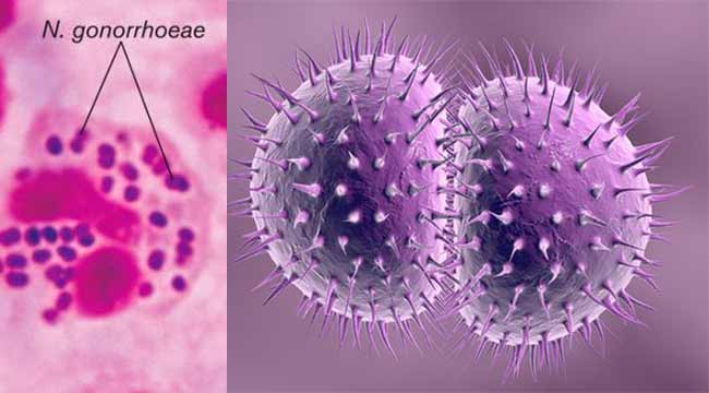
Gonorrhea is a bacterial infection caused by Neisseria gonorrhoeae, a gram-negative diplococcus. Its structure enables infection through specific surface features and mechanisms. Neisseria gonorrhoeae appears as paired kidney bean-shaped bacteria under a microscope. It is an obligate human pathogen that thrives on mucosal surfaces.
📌 Key Takeaways: Gonorrhea Structure & Behavior
- Bacterial Form: Neisseria gonorrhoeae is a Gram-negative, non-encapsulated diplococcus that characteristically appears as paired, kidney bean-shaped cells under a microscope.
- Attachment Mechanism: The bacterium relies heavily on Type IV pili to attach firmly to host epithelial cells, facilitate twitching motility, and exchange genetic material.
- Invasion & Survival: Opa (Opacity-associated) proteins bind to host receptors to trigger cell invasion, allowing the bacteria to multiply both extracellularly and intracellularly.
- Immune Evasion: It constantly alters its surface proteins via antigenic and phase variation to avoid detection by the host immune system and survive inside neutrophils.
The Cell Envelope
The bacteria has a typical gram-negative structure: a thin peptidoglycan layer between inner cytoplasmic and outer membranes. The outer membrane contains lipooligosaccharide (LOS), an endotoxin triggering inflammation, and porin proteins like PorB for nutrient uptake and immune evasion.
Surface Structure Components
Hair-like type IV pili cover the surface, aiding attachment to host cells, twitching motility, and DNA uptake for genetic variation. Opacity-associated (Opa) proteins promote invasion by binding host receptors like CEACAMs.
Resistance systems like the Mtr efflux pump (MtrCDE) expel antibiotics; its MtrF component forms a bowl-shaped dimer with nine transmembrane helices and hairpins, creating a substrate tunnel. Pili and Opa undergo antigenic variation to dodge immunity.
What Is the Life Cycle of Gonorrhea?

Gonorrhea does not have a complex life cycle like parasites; it’s caused by the bacterium Neisseria gonorrhoeae, which replicates directly on human mucosal surfaces through extracellular growth and invasion. The infection process involves attachment, colonization, immune evasion, and potential dissemination.
The Stages of Infection
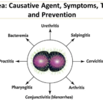
The bacterium first attaches to epithelial cells using type IV pili and Opa proteins, forming microcolonies on mucosal surfaces like the urethra, cervix, rectum, or pharynx. It then invades by endocytosis, transcytoses through cells, and exits into subepithelial spaces, reaching densities of 10^7-10^10 CFU/ml in days.
How Does Neisseria gonorrhoeae Evade the Immune System?
N. gonorrhoeae undergoes phase and antigenic variation in pili and Opa proteins to dodge antibodies and phagocytosis.
It survives inside neutrophils by resisting oxidative bursts and manipulating cytokine responses.
Efflux pumps like MtrCDE expel antimicrobials.
What Is the General Morphology of Gonorrhea Bacteria?
Neisseria gonorrhoeae, the bacteria causing gonorrhea, is a gram-negative diplococcus with distinct morphological features. It appears as paired, kidney bean- or coffee bean-shaped cocci, typically observed inside neutrophils in clinical samples.
Basic Shape and Size
The bacteria are spherical cocci measuring 0.6 to 1.0 micrometer in diameter. They arrange in pairs (diplococci) with flattened adjacent sides, giving the characteristic “kidney bean” profile under Gram staining.
Grammatical and structural Traits
Gram-negative due to a thin peptidoglycan layer. It stains pink/red. Non-encapsulated, non-spore-forming, and aerobic, it shows no true motility but exhibits twitching via type IV pili.