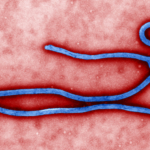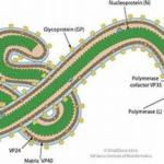


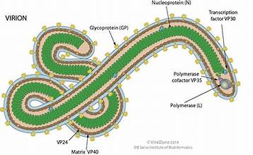
Structure of Ebola Virus – The Ebola virus is a filamentous, enveloped virus with a thread-like or sometimes U-, 6-, or circular shape. Its outer membrane has glycoprotein spikes that help it attach to and enter host cells, and inside it contains a matrix layer and a nucleocapsid with its RNA genome.
📌 Key Takeaways: Ebola Virus Structure
- Virion Shape: The Ebola virus belongs to the Filoviridae family, characterized by a long, enveloped, filamentous (thread-like) structure that can appear flexible, coiled, or shaped like a “U” or “6”.
- Key Surface Proteins: The viral envelope is studded with surface Glycoprotein (GP) spikes, which are crucial for attaching to and entering host target cells.
- The Matrix Layer: Beneath the lipid membrane lies a structural matrix layer composed primarily of VP40 (driving assembly and budding) and VP24 (supporting particle formation and blocking host immune defenses).
- Genomic Core: The core houses a nucleocapsid enclosing a single-stranded, negative-sense, non-segmented RNA genome that codes for seven major structural proteins.
What Is the Overall Structure of the Ebola Virus?
- It belongs to the family Filoviridae, so it has a long, filament-like appearance
- The virion is roughly 80 nm in diameter and about 970 nm long.
- It has a lipid envelope derived from the host cell membrane
- Surface glycoprotein spikes project from the envelope and are important for cell entry.
- Beneath the envelope is a matrix layer made mainly of viral proteins such as VP40 and VP24
- The core contains a nucleocapsid that surrounds a single-stranded, negative-sense RNA genome.
- The genome is non-segmented and encodes seven major structural proteins, including NP, VP35, VP40, GP, VP30, VP24, and L.
It means that the Ebola virus is an enveloped, filamentous, negative-sense ssRNA virus with glycoprotein spikes, a matrix protein layer, and a nucleocapsid enclosing its genome.
How Do VP40 and VP24 Proteins Function During Infection?
VP40 mainly drives virus assembly and budding, helping new Ebola virions form at the host cell membrane and leave infected cells.
VP24 helps with nucleocapsid organization and also blocks parts of the host’s interferon response, which weakens the immune defense during infection.
The Role of the VP40 Matrix Protein
- Acts as the major matrix protein of Ebola virus.
- Coordinates assembly of viral components at the cell membrane
- Promotes budding,so newly made virions can exit the infected cell
- Also contributes to the filamentous shape of the virion
The Role of the VP24 Matrix Protein
- Functions as a minor matrix protein
- Interacts with nucleocapsid components to support proper particle formation.
- Helps Ebola evade the immune system by antagonizing interferon signaling
Thus, VP40 is mainly for assembly and budding, while VP24 helps with particle formation and immune evasion.
What Is the Mechanism of Ebola Virus Cell Entry?
The Ebola virus enters host cells through attachment, uptake, and membrane fusion. Its surface glycoprotein binds to cell-surface factors, the virus is taken into endosomes, and after processing in acidic compartments, it uses the host receptor NPC1 to trigger fusion and release its genome into the cytoplasm.
Step-by-Step Ebola Internalization Pathway
- Attachment: The viral glycoprotein GP helps Ebola bind to host-cell surface factors
- Internalization: The virus is taken into the cell by endocytosis and transported into endosomes/lysosomes
- Processing: Host enzymes in the endosomal compartment cleave GP, which is needed for the next step of entry.
- NPC1 binding: The processed virus binds to the host protein NPC1, a key trigger for infection
- Fusion and release: The viral envelope fuses with the endosomal membrane, and the viral RNA is released into the cytoplasm to begin replication.
Hence, Ebola enters cells by GP-mediated endocytosis, NPC-dependent fusion, and genome release into the cytoplasm.
Which Host Enzymes Trigger Glycoprotein Cleavage?
The main host proteases involved in Ebola GP cleavage are cathepsins in the endosome, especially cathepsin B and cathepsin L. In some contexts, furin-like proteases may also be involved in processing viral glycoproteins, but for Ebola entry, the key cleavage step is usually attributed to endosomal cathepsins.
How Endosomal Cathepsins Activate the Virus
- Cathepsin B and cathepsin L trim GP inside acidic endosomes, which helps expose the site needed forNPC1 binding
- This cleavage makes the glycoprotein competent for membrane fusion and entry.
- Furin is a host protease that cleaves many viral glycoproteins, but it is not the main enzyme class usually emphasized for Ebola GP activation during entry.
Endosomal vs. Surface Proteases in Viral Entry
Endosomal proteases act after the virus is taken into the cell, usually inside acidic endosomes or lysosomes, where they cleave viral proteins to activate membrane fusion and uncoating. Surface proteases act at or near the cell membrane, activating viral glycoproteins before or during entry, so the virus can fuse directly at the cell surface or become entry-competent sooner.
Key Structural Differences at a Glance
- Location: Endosomal proteases work inside endocytic compartments; surface proteases work on the plasma membrane or extracellular side.
- Timing: Endosomal cleavage happens after internazation; surface cleavage happens before or during entry at the cell surface.
- Effect: Endosomal proteases usually trigger a later fusion step in acidic compartments, while surface proteases can prime viral proteins for immediate entry.
- pH Environment: surface proteases are at Neutral pH (~7.4), while endosomal proteases are at low acidic pH (4.5 to 6.0)
- Entry Speed: surface proteases have a fast entry route, while endosomal proteases is slower entry route (complex trafficking)
- Major Examples: Surface protease is TMPRSS2, while cathepsin (B, L) CPL (cathepsin-like proteases)
- Viral pathway: In surface proteases, it is direct cell-surface fusion, while the endocytic pathway is via endosomes and lysosomes.
- Therapeutic Target: In surface proteases, TMPRSS2 inhibitors for SARS-CoV-2; in endosomal proteases, lysosomotropic agents and cathepsin inhibitors.
- TMPRSS2 present: Virus uses surface protease pathway (fast entry, lung/intestine infection)
- TMPRSS2 absent: Virus uses endolysosomal pathway (slow, requires acidic pH for cathepsin activation)
For Ebola, the important cleavage step is mainly endosomal, because host cathepsins process GP inside endosomes before NPC1-mediated fusion occurs. In contrast, some other viruses, such as influenza or coronaviruses, may rely more heavily on surface serine proteases for entry activation.
How Do Lysosomotropic Agents Impact Viral Entry?
Lysosomotropic agents inhibit endolysosomal viral entry by raising endosomal/lysosomal pH, preventing acidification-dependent protease activation and membrane fusion. These compounds accumulate in lysosomes and neutralize the acidic environment required for viral glycoprotein cleavage.