
Life Cycle
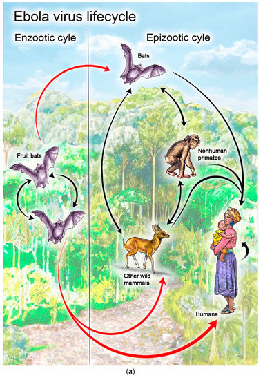
The Ebola virus has a cytoplasmic replication cycle: it attaches to a host cell, enters by endocytosis/macropinocytosis, releases its RNA into the cytoplasm, makes viral mRNA and genome copies, assembles new virions, and buds from the cell surface. Ebola virus has two main phases in its life cycle:
The enzootic cycle in the natural reservoir host (mainly fruit bats) and the epizootic/human cycle after spillover into animals or humans. In the host cell, the virus enters by macropinocytosis, releases its RNA into the cytoplasm, makes viral proteins and genomes, and then assembles and buds from the cell surface.
Intermediate host
The intermediate host is usually a wild animal such as a non-human primate, antelope, or porcupine, which becomes infected after contact with the reservoir host or contaminated material. In these animals, Ebola replicates in the same general way as in other susceptible cells: attachment, entry, cytoplasmic replication, assembly, and release of new virions. These animals can then transmit the virus to humans through handling or contact with infected tissues, blood, or carcasses.
Definitive host
For Ebola, the natural reservoir/definitive host is thought to be fruit bats of the Pteropodidae family. In this host, the virus is maintained in nature and circulates without necessarily causing the severe disease seen in humans and other primates. The viral life cycle inside bat cells still follows the same basic intracellular steps: entry, RNA synthesis in the cytoplasm, protein production, assembly, and budding.
Simple flow:
- Bat reservoir host: virus is maintained in nature.
- Spillover to intermediate animal host: infection of wild mammals such as primates or antelope.
- Spillover to humans: direct contact with infected animals or fluids.
- Human-to-human spread: by contact with infected blood or body fluids.
- Natural host: fruit bats; intermediate host: infected wild mammals; inside each host cell: attachment → entry → RNA → replication → assembly → budding.
What Is the Step-by-Step Ebola Virus Life Cycle?
- Attachment: Viral surface glycoprotein binds host cell receptors and attachment factors such as DC-SIGN/DC-SIGNR and TIM1/HAVCR1 Viral surface glycoprotein binds host cell receptors and attachment factors following entry pathways documented by the National Institutes of Health (NIH).
- Entry: The virus enters the cell mainly by macropinocytosis or endocytosis.
- Endosomal processing: In the endosome, host proteases such as cathepsins help process the glycoproteins, and low pH helps trigger membrane fusion.
- Fusion and uncoating: The viral envelope fuses with the endosomal membrane, releasing the ribonucleocapsid into the cytoplasm.
- Transcription: The viral RNA-dependent RNA polymerase transcribes the negative-sense RNA genome into capped and polyadenylated mRNAs
- Protein synthesis: Host ribosomes translate these mRNAs into viral proteins in the cytoplasm and on the ER
- Genome replication: Once enough nucleoproteins is present, the virus switches from mRNA production to full genome replication through a positive-sense antigenome intermediate.
- Assembly: Viral genomes and structural proteins gather at the plasma membrane to form new nucleocapsids
- Budding and release: New virions bud from the cell membrane, acquiring their envelope as they leave the cell
- Important Features: Ebola is a negative-sense single-stranded RNA virus, so it must carry its own polymerase to start replication. Replication occurs in the cytoplasm, not the nucleus. The virus uses host factors and specialized intracellular compartments to organize RNA synthesis and assembly.
Thus, Ebola is a virus that gets into the cell, copies its RNA in the cytoplasm, makes proteins, builds new particles, and then exits by budding.
How Does the Ebola Virus Evade the Immune System in Fruit Bats?
In fruit bats, Ebola does not seem to “win” by completely hiding; instead, bats appear to control the virus with a very different immune balance that limits damage while keeping the virus in check. Studies suggest bats may have some antiviral defenses that are already active at baseline, which helps suppress viral replication without triggering the strong inflammatory response seen in humans
What helps bats tolerate Ebola:
- Bats appear to have parts of their innate immune system constantly switched on, so they can restrain viruses early.
- This may reduce the need for a large, damaging inflammatory reaction, allowing bats to coexist with viruses that are deadly in other animals.
- Researchers also think bat immune genes are involved rapidly, which may help them handle persistent viral exposure.
How Ebola behaves differently:
- In humans, Ebola can suppress early immune responses, which allows rapid replication before antibodies rise.
- In fruit bats, the virus may still replicate, but the bat immune system seems better at limiting the spread and avoiding harmful overreaction.
- Current evidence supports a host-tolerance model in bats more than a complete immune escape model by the virus.
- Ebola in fruit bats is thought to be controlled by a bat immune system that is more “pre-alert” and less inflammatory, so the bat can carry the virus without severe disease. That is one reason fruit bats are considered a natural reservoir for Ebola-related viruses.
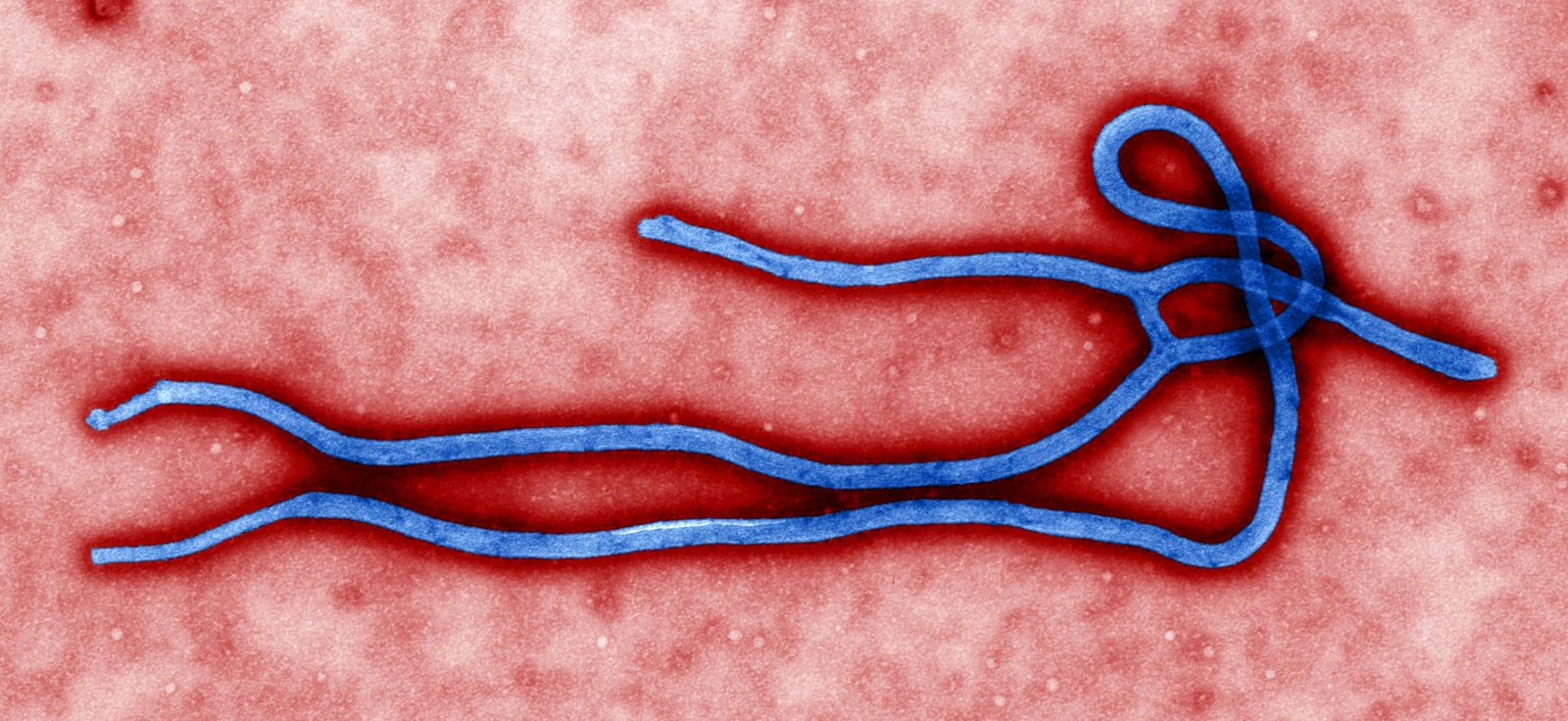
What Is the Morphology of the Ebola Virus?
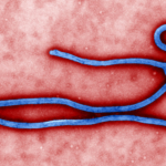
The Ebola virus has distinctive filamentous (thread-like) morphology, which is why it belongs to the family Filoviridae (from Latin filum = thread).
Morphological features:
- Shape: It is long, cylindrical/tubular filaments; it can also appear “U-shaped,” circular, or like a “6” (shepherd’s crook)
- Diameter: Consistently ~ 80 nm (nanometers)
- Length: Typically ~ 970 nm, but can range up to 14,000 nm; highly variable and pleomorphic
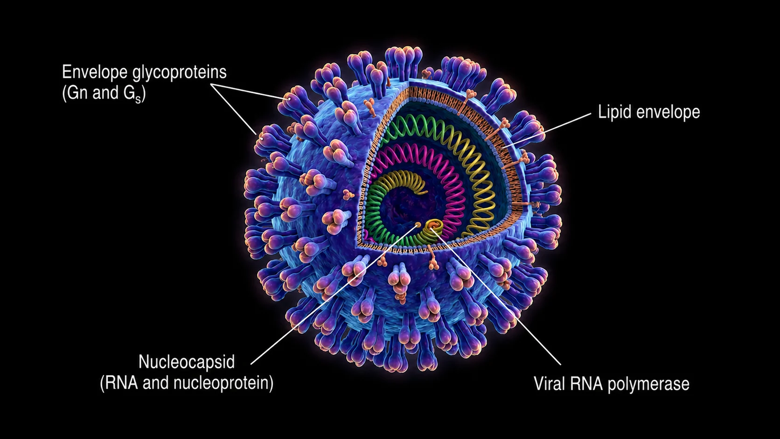
- Surface: Lipid bilayer envelope with 7 to 10 nm glycoprotein (GP) spikes projecting outward, spaced ~ 10 nm apart
- Internal structure: Contains viral envelope, matrix (VP40 proteins), and nucleocapsid (RNA genome wrapped in nucleoprotein)
- Genome: Non-segmented, single-stranded negative-sense RNA with 7 genes.

The virus is pleomorphic (exists in many shapes) and appears flexible with varying degrees of twisting. Electron microscopy shows it resembles “a length of thread” or “bowl of spaghetti” when filaments fuse end to end