


Here is a chronological history of the filarial disease
Ancient Times
Evidence of filariasis (specifically elephantiasis) is seen in ancient Egypt, depicted by swollen limbs.
6th Century BC
Filariasis was described by the famous Indian physician Susruta in his book, “Susruta Samhita.”
7th Century AD
Madhavkara described signs and symptoms of the filarial disease in his treatise “Madhava-Nidhana,” which are still considered valid today.
1588–1592
The Dutch merchant Jan Huyghen-Van Linschoten described an elephantiasis-like disease in Goa, India.
1709
Clarke called elephantoid legs in Cochin ‘Malabar legs.’
1863
Demarquay in Paris found microfilariae in hydrocele fluid.
1866
Wucherer discovered microfilariae in chyluria.
1872
Lewis found microfilariae in peripheral blood in Calcutta.
1876
Erwin Von Baelz detected microfilariae in the blood in Tokyo.
1877
Bancrofti found an adult female filarial worm in a lymph node ulcer.
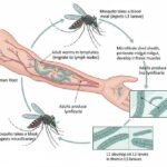
Manson discovered microfilariae in mosquitoes, establishing the mosquito as the vector.
1879
Manson described the nocturnal periodicity of microfilariae in the blood.
1888
Sibthorpe identified adult male worms.
Early 20th Century
Filariasis was widely reported in tropical and subtropical countries.
1960
The species from Timor-Leste was classified as Brugia timori
Definition and Impact of Filariasis
Filariasis is a group of diseases caused by parasitic nematodes (worms) and transmitted through mosquitoes or flies. Signs and symptoms include swelling (or hydrocele) of the lower and upper limbs. Filariasis leads to irreversible chronic manifestations that cause social stigma, considerable economic loss, and severe physical disability to affected individuals. Acute attacks of filariasis frequently traumatize patients with transient episodes of disability, often requiring bed rest for a few days.
Kinds of Filarial Parasites
There are three main groups of filarial parasites and a total of eight species known to cause filariasis in humans. They are mainly classified based on their habitat and species. These filariae belong to the superfamily ‘Filarioidea’ and are transmitted by various insects like mosquitoes and flies.
A) Lymphatic Filariasis
This group includes three species:
- Wuchereria bancrofti
- Brugia malayi
- Brugia timori
B) Non-Lymphatic Filariasis
This group is divided into two subgroups:
- Cutaneous group
- Body cavity group
C) Cutaneous Group
This group includes two species:
- Loa loa (or Loiasis)
- Onchocerca volvulus (River Blindness)
D) Body Cavity Group
There are three species in this group:
- Mansonella perstans (MP)
- Mansonella ozzardi
- Mansonella streptocirca cerca
Zoonotic Parasites
Additionally, some species of the genus Dirofilaria can occasionally infect humans as zoonotic parasites, though they are primarily parasites of animals. Examples include Dirofilaria immitis (heartworm) and Dirofilaria repens.