


Recognition and Features of Malaria Parasites
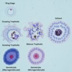
Malaria parasites take up JSB stain in a special way in both thick and thin blood films. You must be able to distinguish the various parts of the malaria parasites, or general features of Plasmodium, as shown in the diagram that follows.
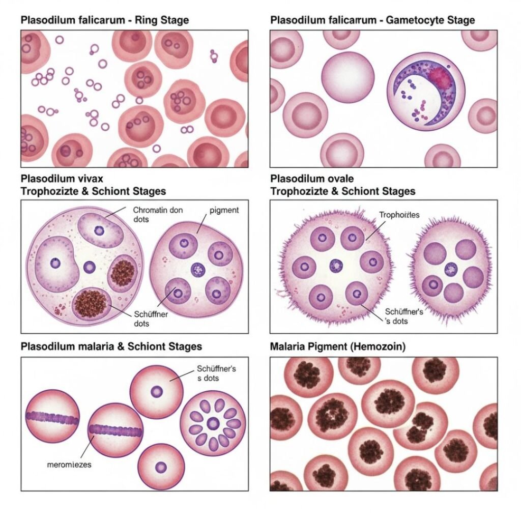
Malaria parasites pass through a number of developmental stages. In all stages, however, the same parts of the parasite will stain the same colour.
The key parts of the malaria parasite are
The key parts of the malaria parasite are
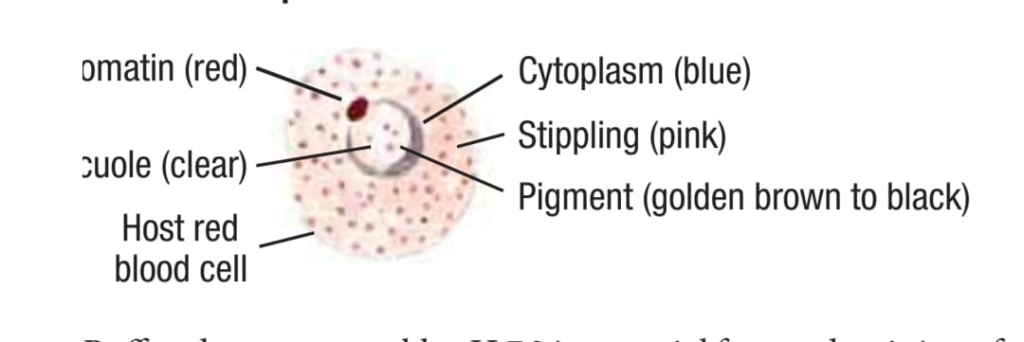
1) Chromatin or Chromatin Dots (Nucleus)
The chromatin dots are the nucleus of the parasite. It is the respiratory organ of the parasite. It is usually round in shape and stains deep red.
2) Cytoplasm
Cytoplasm occurs in a number of forms, from a ring shape to a totally irregular shape. It always stains blue in colour, although the shade of blue may vary between the malaria species.
3) Pigment
Pigment appears as the parasite grows. Malaria pigment is a by-product of the growth or metabolism of the parasites. During the growth of Plasmodium parasites inside the red blood cells, the parasite digests hemoglobin, which produces a large amount of waste product called hemozoin, or pigment.
- Hemozoin is a combination of hematin and protein.
- The size, shape, and colour of hemozoin, or pigment, vary depending on the parasite species and developmental stage.
- It does not take up stain but has a colour of its own, which may range from pale yellow to dark brown or black pigmented granules in the cytoplasm of the parasite.
- These granules are small, angular, or rod-like and increase in number with the growth of the parasite.
4) Vacuole
The vacuole is the digestive organ of the parasite. This lysosome-like organelle is essential for the parasite’s survival during its blood stage development. The vacuoles are specialized compartments for malaria parasites, which protect the parasites from the host defense mechanism and allow them to regulate the exchange of nutrients, waste, and proteins between the parasite and the host cell cytoplasm.
5) Stippling
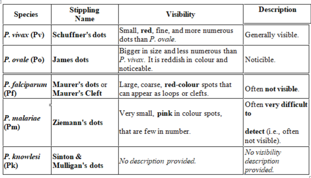
Stippling refers to the fine dots or granules that appear on the red blood cell (RBC) infected with malaria parasites. Stippling is often described as the “footprint” of the parasite. It is one of the key morphological features used by microscopists to identify the Plasmodium species in a thin blood smear. The visibility of stippling depends heavily on the preparation of the blood sample and the pH of the stain used.
Types of Stippling and Associated Species
Mechanism of Stippling
The parasite exports proteins to the red blood cell membrane to modify it, which increases the host cell’s permeability for nutrients and helps the parasite grow.