Treponema pallidum, the bacterium causing syphilis, has a distinctive helical spirochete structure adapted for motility and host invasion.
Structure of Treponema pallidum
Cell Morphology
It appears as a thin 0.1 to 0.2 μm diameter, flexible, coiled rod 6 to 20 μm long with 6 to 14 regular spirals; it is non-motile outside the host but highly motile via flagella in vivo.
Envelope Layers
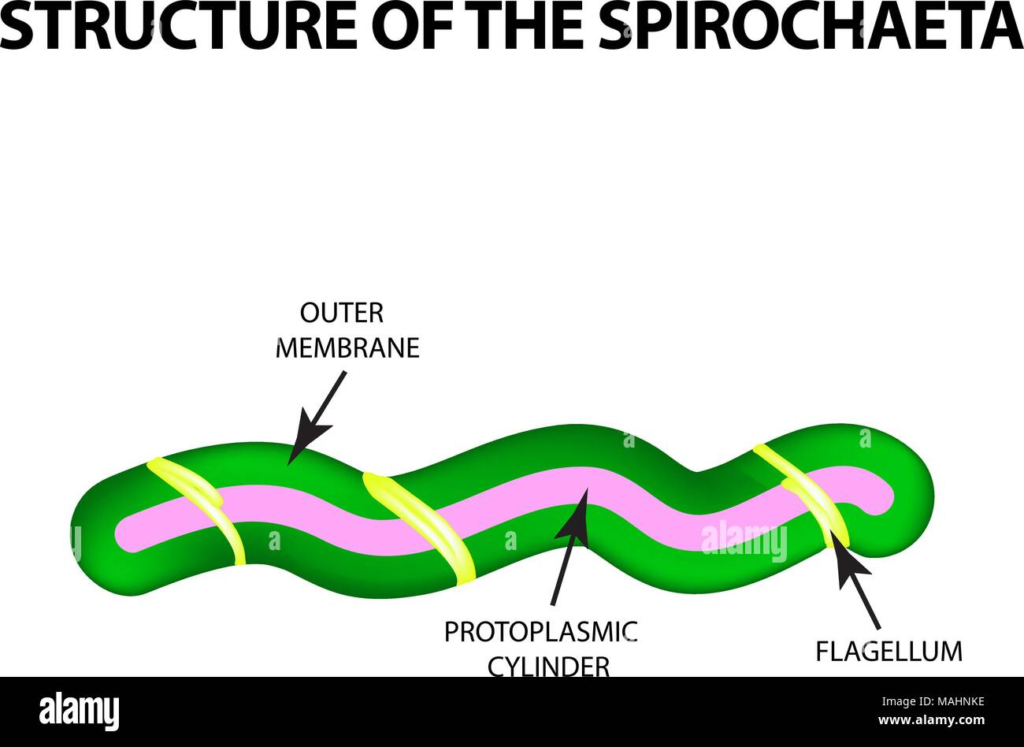
1) Outer membrane
Lacks LPS (unlike typical Gram-negatives); contains lipoproteins, few OMPs (e.g., TPO126, TPO479 beta-barrels), and Tpr proteins for immune evasion.
2) Periplasmic space
Houses periplasmic flagella (endoflagella) that wrap around the cell body, enabling corkscrew motility.
A thin peptidoglycan layer and inner cytoplasmic membrane complete the Gram-negative-like architecture.
3) Composition
Dry weight ~70% proteins, including adhesins like Tp0751; 20% lipids (phospholipids, cholesterol from host); 5 to 10% carbs; flagella anchored at poles.
Life Cycle of Syphilis
Syphilis progresses through distinct clinical stages driven by Treponema pallidum dissemination in the host, rather than a traditional microbial life cycle with replication cycles.
Incubation Period
Bacteria multiply locally at the entry site 10 to 90 days, averaging 21 days without symptoms, preparing for primary invasion.
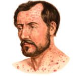
Primary Stage
A chancre (painless ulcer) forms at the mucocutaneous site. Treponemes peak locally, then enter lymphatics/blood for systemic spread in 3 to 6 weeks.
Secondary Stage
Disseminated infection causes rash, fever, and lymphadenopathy 4 to 10 weeks; bacteria are widespread in skin/mucosa and highly infectious.
Latent Stage
Asymptomatic: early latent (< 1–2 years) infectious, late latent (years–decades, less so)
Treponemes persist in tissues.
Tertiary Stage
Gummas, cardiovascular/neurosyphilis in 15%–30% untreated cases 3 to 15+ years, and destructive inflammation from an immune response.
Morphology of Syphilis
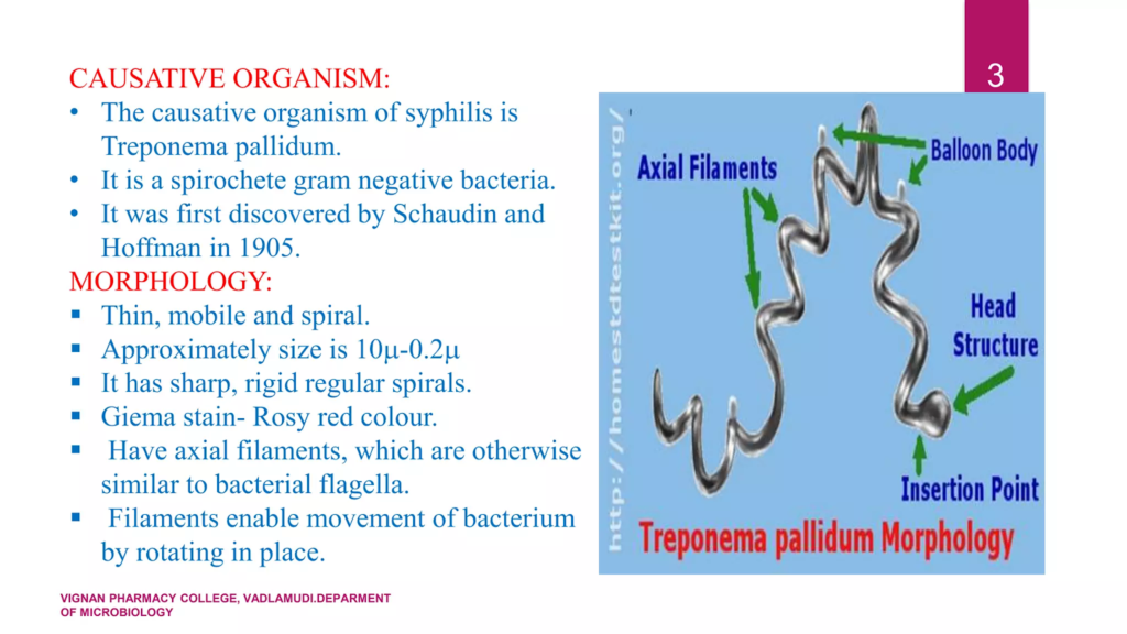
Treponema pallidum, the spirochete bacterium causing syphilis, exhibits a distinctive helical morphology enabling its motility and tissue invasion.
Cell-Shape Dimension
Helically coiled rods, 6 to 20 μm long and 0.1 to 0.2 μm wide, with 6 to 14 regular spirals; flexible, corkscrew-like form for serpentine movement.
Flagellar Structure
Periplasmic (endoflagellar) flagella, 2 at each pole, wrap around the protoplasmic cylinder within the periplasmic cylinder within the periplasmic space; the core of FlaB proteins is sheathed by FlaA, providing torque for rotation.
Envelope Composition
Gram-negative-like: outer membrane lipoproteins, Tpr porins, no LPS, thin peptidoglycan layer, cytoplasmic membrane; cone-shaped tip structures aid attachment.
How does motility of Treponema pallidum contribute to pathogenesis
Treponema pallidum motility, powered by periplasmic flagella enabling corkscrew rotation, is a key virulence factor in syphilis pathogenesis.
Tissue Penetration
High translational and rotational speeds up to 2 revolutions/sec. allow navigation through viscous mucus, gels, and extracellular matrices that impede other bacteria, facilitating initial mucocutaneous entry and deep tissue invasion.
Systemic Dissemination
Motility aids rapid hematogenous spread from the primary chancre via lymph/blood within hours-days, crossing endothelial barriers e.g., blood-brain and placental, despite shear stress; adhesins like Tp0751 slow movement for stable attachment before extravasation.
Immune Evasion and Persistence
Dynamic swimming evades phagocytosis/microbicidal peptides; altered kinematics near host cells promote cytokine induction, barrier disruption, and ECM degradation, enabling chronic latency.
seizures or death shortly after birth; early prenatal screening and penicillin treatment prevent most harm.