Morphology and Characteristics of Plasmodium knowlesi
The parasite Plasmodium knowlesi was first described by the Italian physician Giuseppe Franchini in 1927. He noted that this parasite was distinct from others found in the blood of the long-tailed macaque. The parasite was clearly observed again in 1931 in a long-tailed macaque by H.G.M. Campbell in Calcutta, India.
In 1932, Biraj Mohan Das Gupta and Robert Knowles described its morphology and demonstrated that it could infect humans. The parasite was named P. knowlesi by John Sinton and H. W. Mulligan in 1932.
Features in Red Blood Cells (RBCs)
- Infected RBCs are not enlarged.
- Multiple parasites can be found within a single RBC.
- In P. knowlesi, stippling is known as “Sinton & Mulligan’s.”
- Pigment appears as coarse, dark grains scattered irregularly in the cytoplasm.
The 24-Hour Erythrocytic Cycle
The parasite of P. knowlesi completes its development within 24 hours in the RBC and releases new merozoites every 24 hours. This rapid cycle has several important implications:
- It allows for very rapid multiplication, resulting in a high level of parasitemia in a short time.
- High parasitemia increases the risk of severe malaria.
- The bursting of RBCs every 24 hours causes daily fever cycles, which is called “quotidian fever.”
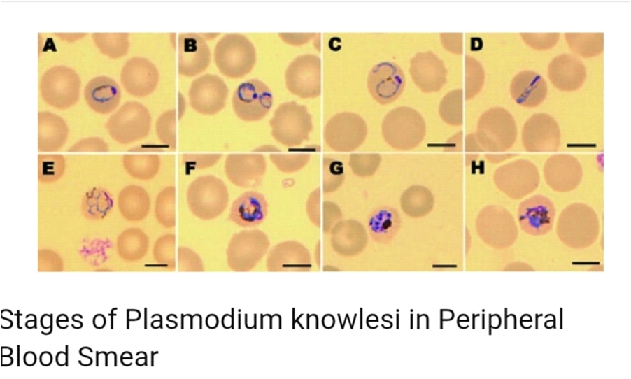
Stages Found in Peripheral Blood Smears
There are mainly four stages found in peripheral blood smears:
1. Ring Stage
- Ring stage forms resemble those of P. falciparum (Pf), often showing double chromatin dots.
- The parasite may appear on the edge of the red blood cell, a characteristic known as the “appliqué” or “accolé” form, which is also seen in P. falciparum.
2. Trophozoite Stage
- Late and mature trophozoites of P. knowlesi closely resemble those of P. malariae (Pm), including band-form trophozoites.
3. Schizont Stage
- P. Knowlesi schizonts contain up to 16 merozoites.
- This is typically more than the 12 merozoites seen in P. malariae (Pm).
4. Gametocyte Stage
- Gametocytes are spherical, filling the host RBC with pale cytoplasm and a darker eccentric nucleus.
Differentiating P. knowlesi
P. Knowlesi can be difficult to differentiate from other Plasmodium species by microscopy due to morphological similarities.